Science

Science and Research

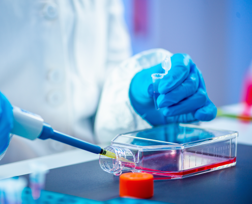
Non-growth-based methods for the phenotypic investigation of microorganisms offer fast and label-free direct access to identity determination, vital metabolic functions and the investigation of microbial interactions with substances. Academic applications of the software-controlled, fully automated modular instrument concept GramRay.

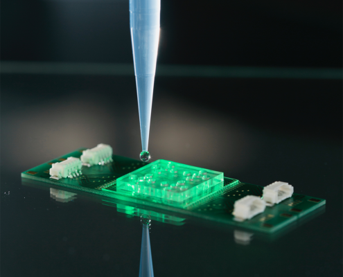
Raman spectroscopy differs from other currently used techniques by its easy application at low cost, its high analysis speed and its broad information content both about the chemical composition and the structure of biomolecules within microorganisms.

Slight changes in the chemical composition of microorganisms can be monitored by Raman spectroscopy and used to differentiate genera, species or even strains. Pathogens can be detected from complex matrices such as soil, food and body fluids. In addition, spectroscopic studies of host-pathogen interactions and the effect of antibiotics on bacteria are also dealt with.

Academic applications use the highest sensitivity in single cell identification for the investigation of zoonoses and the detection of pathogens from beverages and food. Vibrions such as cholera are living, difficult-to-cultivate germs (VBNC) that can be reliably detected with GramRay without a previous cultivation step. Their metabolism is investigated with stable isotopes Raman (SIRM). In the elucidation of microbiomes, GramRay provides direct access to populations, as growth and incubation steps are not required. Raman also shows non PCR primer specific gaps, such as the widely used Next Generation Sequencing method for the elucidation of the microbiome.

With the GramRay system, the viability of bacteria can be examined directly. Interactions with substances or host-pathogen interactions are immediately visible. Thus the suitability of potential antibiotics for specific bacteria can be determined within only 1 hour. By visualizing the interaction of an antibiotic with the bacterial membrane, the mechanisms of antibiotic resistance are elucidated.

    Features & Benefits

    • Sample volume: 0.1 – 500 ml
    • Working time for the laboratory technician: 10-30 min
    • Sensitivity: 10 microbes depending on the matrix and separation method
    • Time to result: 90 min
    • Counting of the total viability count: 15 minutes
    • Time for genus/species classification: 15 s per microbe
    • Integrated proven software for modeling specific chemometric and machine learning methods:
      • linear discriminant analysis (LDA)
        Random Forest (RF)
        k-nearest neighbors (kNN)
      • Support Vector Machine (SVM)
        partial least squares of discriminant analysis (PLS-DA)
        unsupervised
      • hierarchical Ward-Cluster-Analysis (HCA)
      • linear regression; partial least squares of regression (PLSR).
      • Integrated validation algorithms:
        • leave-one-batch-out cross-validation (LOBOCV)
        • 10-fold cross-validation
    • Flexible sample quantity
      Efficient work and fast preparation of many samples
    • Automation increases the reproducibility of research results
    • Highest sensitivity and accuracy: every living cell is counted
    • differentiation between:
      • dead/living/VBNC
    • High sample throughput and spectra generation for modeling
    • User hierarchy and data integrity enables traceability of test results

    Part of the Publication list:

    • S. Stöckel, S. Meisel, R. Böhme, M. Elschner, P. Rösch and J. Popp, ” Effect of supplementary manganese on the sporulation of Bacillus endospores analysed by Raman spectroscopy”, J. Raman Spectrosc. 2009, 40, 1469-1477.
    • S. Meisel, S. Stöckel, M. Elschner, P. Rösch and J. Popp, “Assessment of two isolation techniques for bacteria in milk towards their compatibility to Raman spectroscopy”, Analyst 2011, 136, 4997-5005.
    • S. Stöckel, S. Meisel, M. Elschner, P. Rösch and J. Popp, “Raman Spectroscopic Detection of Anthrax Endospores in Powder Samples”, Angew. Chem. Int. Ed. 2012, 51, 5339-5342.
    • S. Meisel, S. Stöckel, M. Elschner, F. Melzer, P. Rösch and J. Popp, “Raman spectroscopy as a potential tool for the detection of Brucella spp. in milk”, Appl. Environ. Microbiol. 2012, 78, 5575-5583.
    • S. Stöckel, S. Meisel, M. Elschner, P. Rösch and J. Popp, “Identification of Bacillus anthracis via Raman spectroscopy and chemometric approaches”, Anal. Chem., 2012, 84, 9873−9880.
    • U. Münchberg, L. Wagner, E. T. Spielberg, K. Voigt, P. Rösch and J. Popp, “Spatially resolved investigation of the oil composition in single intact hyphae of Mortierella spp. with Raman micro-spectroscopy”, Biochim. Biophys. Acta Mol. Cell Biol. Lipids, 2013, 1831, 341-349.
    • S. Pahlow, S. Kloß, K. Kirsch, U. Hübner, P. Rösch, K. Weber and J. Popp, “Isolation and Identification of Pathogens with a Surface Modified Aluminium-Chip”, ChemPhysChem, 2013, 14, 3600-3605.
    • S. Kloß, B. Kampe, S. Sachse, P. Rösch, E. Straube, W. Pfister, M. Kiehntopf and J. Popp, ” Culture independent Raman spectroscopic identification of urinary tract infection pathogens – A proof of principle study”, Anal. Chem., 2013, 85, 9610-9616.
    • S. Meisel, S. Stöckel, P. Rösch and J. Popp, “Identification of meat associated pathogens via Raman microspectroscopy”, Food Microbiol. 2014, 38, 36-43.
      D. Kusić, B. Kampe, P. Rösch and J. Popp, “Identification of water pathogens by Raman microspectroscopy”, Water Res., 2014, 48, 179-189.
    • E. Kämmer, K. Olschewski, T. Bocklitz, P. Rösch, K. Weber, D. Cialla and J. Popp, “A new calibration concept for a reproducible quantitative detection based on SERS measurements in a microfluidic device demonstrated on the model analyte adenine”, PCCP 2014, 16, 9056-9063.
    • U. Münchberg, P. Rösch, M. Bauer and J. Popp, “Raman spectroscopic identification of single bacterial cells under antibiotic influence”, Anal. Bioanal. Chem. 2014, 406, 3041-3050.
    • A. Silge, W. Schumacher, P. Rösch, P. A. da Costa Filho, C. Gérard and J. Popp, “Identification of water conditioned Pseudomonas aeruginosa by Raman microspectroscopy on a single cell level”, Syst. Appl. Microbiol. 2014, 37, 360–367.
    • S. Kloß, P. Rösch, W. Pfister, M. Kiehntopf and J. Popp, “Towards culture free Raman spectroscopic identification of pathogens in ascitic fluid”, Anal. Chem. 2015, 87, 937-943.
    • S. Stöckel, S. Meisel, M. Elschner, F. Melzer, P. Rösch and J. Popp, “Raman spectroscopic detection and identification of Burkholderia mallei and Burkholderia pseudomallei in feedstuff”, Anal. Bioanal. Chem. 2015, 407, 787–794.
    • S. Stöckel, J. Kirchhoff, U. Neugebauer, P. Rösch and J. Popp, “The application of Raman spectroscopy for the detection and identification of microorganisms”, Journal of Raman Spectroscopy 2015, 89-109.
    • U. Münchberg, L. Wagner, C. Rohrer, K. Voigt, P. Rösch, G. Jahreis and J. Popp, “Quantitative assessment of the degree of lipid unsaturation in intact Mortierella fungi by Raman microspectroscopy”, Anal. Bioanal. Chem. 2015, 407, 3303–3311.
    • V. Kumar B.N., B. Kampe, P. Rösch and J. Popp, “Characterization of carotenoids in soil bacteria and investigation of their photodegradation by UVA radiation via resonance Raman spectroscopy”, Analyst 2015, 140, 4584-4593.
    • D. Kusić, B. Kampe, A. Ramoji, U. Neugebauer, P. Rösch and J. Popp, “Raman spectroscopic differentiation of planktonic bacteria and biofilms”, Anal. Bioanal. Chem. 2015, 407, 6803–6813.
    • S. Kloß, B. Lorenz, P. Rösch, S. Dees, I. Labugger and J. Popp, “Destruction-free procedure for the isolation of bacteria from sputum samples for Raman spectroscopic analysis”, Anal. Bioanal. Chem. 2015, 407, 8333–8341.
    • S. Stöckel, A. S. Stanca, J. Helbig, P. Rösch and J. Popp, “Raman spectroscopic monitoring of the growth of pigmented and non-pigmented Mycobacteria”, Anal. Bioanal. Chem. 2015, 407, 8919–8923.
    • V. Kumar B.N., B. Kampe, P. Rösch and J. Popp, “Classification and identification of pigmented cocci bacteria relevant to the soil environment via Raman spectroscopy”, Environ. Sci. Pollut. Res. 2015, 22, 19317–19325.
    • S. Pahlow, S. Stöckel, S. Pollok, D. Cialla-May, P. Rösch, K. Weber and J. Popp, “Rapid identification of Pseudomonas spp. via Raman Spectroscopy Using Pyoverdine as Capture Probe”, Anal. Chem. 2016, 88, 1570−1577.
    • D. Kusić, A. Ramoji, U. Neugebauer, P. Rösch and J. Popp, “Raman spectroscopic characterization of packaged L. pneumophila strains expelled by T. thermophila”, Anal. Chem. 2016, 88, 2533-2537.
    • D. Kusić, P. Rösch and J. Popp, “Fast label-free detection of Legionella spp. in biofilms by applying immunomagnetic beads and Raman spectroscopy”, Syst. Appl. Microbiol. 2016, 39, 132–140.
    • V. Kumar B.N., S. Guo, T. Bocklitz, P. Rösch and J. Popp, “Demonstration of carbon catabolite repression in naphthalene degrading soil bacteria via Raman spectroscopy based stable isotope probing”, Anal. Chem. 2016, 88, 7574-7582.
    • S. Stöckel, S. Meisel, B. Lorenz, S. Kloß, S. Henk, S. Dees, E. Richter, S. Andres, M. Merker, I. Labugger, P. Rösch and J. Popp, “Raman spectroscopic identification of Mycobacterium tuberculosis”, J. Biophotonics 2017, 10, 727-734.
    • M. Taubert, S. Stöckel, P. Geesink, S. Girnus, N. Jehmlich, M. von Bergen, P. Rösch, J. Popp and K. Küsel, “Tracking active groundwater microbes with D2O labeling to understand their ecosystem function”, Environ. Microbiol. 2018, 20, 369-384.
    • S. Guo, A. Kohler, B. Zimmermann, R. Heinke, S. Stöckel, P. Rösch, J. Popp and T. W. Bocklitz, “Extended Multiplicative Signal Correction Based Model Transfer for Raman Spectroscopy in Biological Applications”, Anal. Chem. 2018, 90, 9787-9795.
    • N. Ali, S. Girnus, P. Rösch, J. Popp and T. Bocklitz, “Sample size planning for multivariate data: a Raman spectroscopy based example”, Anal. Chem. 2018, 90, 12485-12492.
    • B. Lorenz, P. Rösch and J. Popp, “Isolation matters- Processing blood for Raman microspectroscopic identification of bacteria”, Anal. Bioanal. Chem. 2019, 411, 5445–5454.
    • C. Wichmann, M. Chhallani, T. Bocklitz, P. Rösch and J. Popp, “Simulation of transportation and storage and their influence on Raman spectra to bacteria”, Anal. Chem. 2019, 91, 13688-13694.
    • A. A. Moawad, A. Silge, T. Bocklitz, K. Fischer, P. Rösch, U. Roesler, M. C. Elschner, J. Popp and H. Neubauer, “A machine learning-based Raman spectroscopic assay for the identification of Burkholderia mallei and related species”, Molecules 2019, 24, 4516.